
Some photographs show illness or injuries that readers may find distressing. These images are hidden behind labelled expandable sections, so you can read the article without opening them.
The Octodon degus is a generally robust rodent, and its main disease risks can be reduced by appropriate nutrition and housing. However, this species has some pathologies that are less common in other small pets. Recognising signs of illness helps owners seek veterinary care promptly.
Some of the health problems discussed in this article have already been covered in detail in our health category.
Only a veterinarian familiar with degus is qualified to make a diagnosis and propose appropriate treatments. Be cautious about self-diagnosing your animals, as it can lead to a worsening of their health.
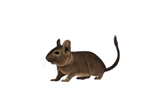
Diseases and health problems in degus
The Octodon degus is a relatively recent pet animal that emerged from the use of the species in laboratories1. Its non-domesticated status and particular husbandry needs make informed care important. Likewise, scientific studies on its health problems are less numerous than for other species like rabbits2. However, in recent years, veterinary publications have helped improve our understanding of this species. The octodon is also often used in laboratories to study diabetes and Alzheimer’s disease-like pathologies, contributing to a better understanding of the diseases that can affect the degu as a pet.
In captivity, the degu is mainly affected by dental diseases and skin diseases, reported in 60% and 35% of animals, respectively, in a retrospective clinical study of 300 degus; these are not general-population prevalence estimates2. Other pathologies affect fewer degus and are sometimes consequences of other health problems3.
Interpreting the tables: these are published sample data, not universal clinical reference intervals. † marks totals that do not agree with the displayed age-group counts; the original figures are retained pending confirmation. ‡ marks an inconsistency already present in the published blood-data table. A confidence interval for a mean is not an individual reference interval. Results must be interpreted by a veterinarian using the sampling and laboratory methods.
| Disease | Degus | Degus two years and older | Degus younger than 2 years | |||
|---|---|---|---|---|---|---|
| N = 300 | % | N = 190 | % | N = 110 | % | |
| Dental pathologies | 180 † | 60 | 144 † | 75.80 | 34 † | 30.91 |
| Skin pathologies | 110 | 36.67 | 56 | 29.47 | 54 | 49.09 |
| Eye diseases | 50 | 16.67 | 32 | 16.84 | 18 | 16.36 |
| Gastrointestinal pathologies | 30 | 10.00 | 17 | 8.95 | 13 | 11.82 |
| Reproductive pathologies | 28 | 9.33 | 20 | 10.53 | 8 | 7.27 |
| Skeletal pathologies | 22 | 7.33 | 14 | 7.37 | 8 | 7.27 |
| Rhinitis | 16 † | 5.33 | 14 † | 7.37 | 4 † | 3.64 |
| Diabetes | 12 | 4 | 10 | 5.26 | 2 | 1.82 |
| Obesity | 12 | 4 | 9 | 4.74 | 3 | 2.73 |
| Ear diseases (otitis) | 5 | 1.67 | 3 | 1.58 | 2 | 1.82 |
| Renal pathologies | 4 | 1.33 | 4 | 2.11 | 0 | 0 |
| Epileptic seizures (unknown etiology) | 2 | 0.67 | 0 | 0 | 2 | 1.82 |
| Hyperthermia | 1 | 0.33 | 1 | 0.53 | 0 | 0 |
| Diagnosis unknown | 14 | 4.67 | 8 | 4.21 | 6 | 5.45 |
| Healthy animals | 38 | 12.67 | 16 | 8.42 | 22 | 20 |
| † Counts reproduced from the historical site table remain inconsistent in the dental and rhinitis rows. Percentages here are arithmetic from the listed counts; the original full paper table has not been inspected. Cases can have multiple diagnoses. | ||||||
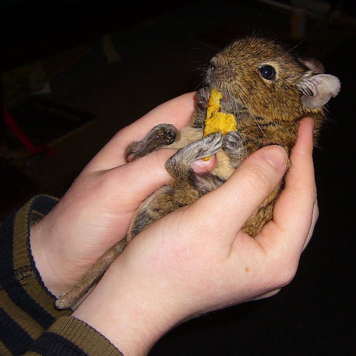
Dental diseases
Dental pathologies, such as malocclusion, affect a high proportion of animals older than two years in a retrospective clinical sample2. This is mainly due to poor nutrition4 5, often low in calcium and fiber. Degus need prolonged chewing opportunities because their teeth grow continuously. In some cases, genetic malocclusions can be observed, but these are rare. Dental problems can develop when teeth wear unevenly or grow out of alignment6. As a result, the teeth become too long and can injure the animal. Some may even grow above or below the tongue, creating “dental bridges.” In severe cases, the teeth can even pierce the skull of the degu. Trauma, such as a fall, can also be the cause of these pathologies. The incisors of degus can also be affected, especially if they grow excessively and then cause mouth injuries.
CT images of the sagittal plane of the maxillary dental arch reconstructed in a 3D model, medio-lateral view (A), and surface reconstruction (B). Apical elongation and the reserve crown of the fourth premolar erupted into the nasal cavity (A), with a bulging apex in the nasal bone (B). Irregular structure suggesting dysplastic changes in dental structures. Reconstructed images are visualized using the Drishti volume rendering software. (Vladimir Jekl and Tomas Zikmund).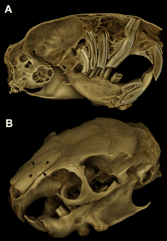
When affected, degus tend to lose weight, have abnormal chewing, and can also develop lung infections7 8. Drooling and refusal to eat are further warning signs requiring examination. The veterinarian can file down overgrown teeth or remove those that are misaligned.
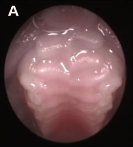
An appropriate fibrous diet and a suitable mineral balance help reduce dental risk, but cannot guarantee prevention. Calcium and phosphorus figures from experimental diets are not universal prescriptions. Provide suitable hay and leafy plant material, and never restrict a painful or underweight animal’s food simply to force hay consumption.
Incisors
Excessive growth
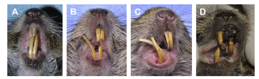
When incisors grow excessively, degus have difficulty eating6 and can injure their mouths and lips. Generally, incisor issues are related to dental problems with molars and premolars. Fractures and dysplasias are sometimes also observed. Dysplasias are tissue lesions and are usually associated with mineral deficiencies or repeated trauma, such as chewing on bars. Incisors can be filed down by a veterinarian, who should check for infection, abscess, or elodontoma.
Incisor problems in degus (Vladimir Jekl). (A) Elongation and depigmentation of enamel on clinical crowns. (B) Severe malocclusion and deviation of the right mandibular incisor after traumatic injury. (C) Iatrogenic right lateral deviation of the mandibular incisor and overall coronal elongation after trimming clinical crowns with pliers. (D) Displacement and malocclusion of incisors secondary to maxillary melanoma.
Whitening / Enamel Issues
Adult degu incisors normally have orange-yellow enamel. A change to pale or white enamel warrants assessment for a possible dietary imbalance or other dental problem9.
Malocclusions of premolars and molars
Malocclusions and pathologies of molars and premolars are very common in degus and are associated with poor nutrition and a lack of natural wear during chewing4 5. Teeth can grow abnormally, either crooked, underneath, or through the mouth, even to the point of perforating the skull. Dental bridges can also form under or over the tongue.
Mazu, a degu with teeth growing through its skull and touching the eyeball, which cannot be operated on.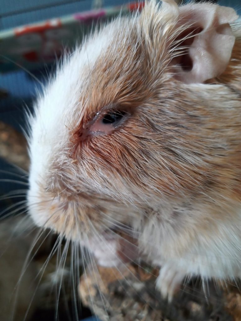
These problems are often silent at first but remain very painful for degus during chewing6 and can lead to elodontoma, rhinitis, and other cranial and ocular pathologies. Cavities and enamel deformations are also noted in degus. Filing or removal of teeth affected by dental pathologies is essential, as well as the implementation of an appropriate diet.
Elodontoma/Odontoma
Elodontomas are proliferative dental lesions in degus10, often linked to malocclusions or other dental pathologies. These lesions are proliferations of dental tissues and can alter the structure of the mouth by shifting dental crowns. Moreover, when elodontomas affect incisors or premolars, they can obstruct the nasal passage and lead to respiratory problems. Treatment depends on the lesion’s location and extent; surgical removal may be considered, but it is a complex surgical procedure that cannot be considered for all animals. If the lesion cannot be treated and suffering cannot be adequately controlled, the veterinarian may discuss euthanasia.
Radiograph of a degu’s skull with dental malocclusion – Photo by Isabelle Wornham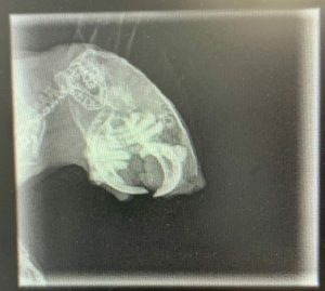
Dental trauma
Dental traumas are often noticeable in incisors, which can be chipped, split, or missing. Generally, this occurs as a result of trauma, such as a fall or severe dental imbalance that weakened the teeth. Stereotypies like continuous bar-biting can also cause dental fractures. It is sometimes necessary to file the opposing tooth to allow normal regrowth of the broken tooth6.
Digestive Pathologies in Degus
Degus can be affected by several digestive pathologies11, with the most well-known being bloat. Digestive disorders were reported in approximately 10% of the animals in the retrospective clinical series and are often linked to poor nutrition12, stress, obesity, or dental problems10. To ensure ideal digestion in degus, their diet must be balanced and contain an adequate amount of fiber.
Gastrointestinal Stasis
Gastrointestinal stasis is a decrease or halt in digestion, sometimes with an accumulation of material in the stomach that is not being emptied. This condition can affect degus, especially after a change in diet, a lack of fiber, or dental problems. Stasis can lead to bacterial overgrowth, colic, and gas accumulation in the stomach.
When affected, degus may show signs of constipation, have an abnormally swollen or “hard” abdomen, refuse to eat, and generally become less active. Since stasis can lead to other issues, degus are at risk, and it is important to immediately consult a veterinarian to attempt to restart the digestive process3 11.
After assessment, the veterinarian may prescribe fluid therapy and a suitable herbivore recovery diet. Suspected obstruction, respiratory distress or impaired swallowing must be addressed before oral syringe feeding.
Intestinal Dysbiosis
Intestinal dysbiosis is an imbalance in the gut flora, leading to the proliferation of enterobacteria (E. coli, Clostridioides difficile). This can result in diarrhea, enteritis, or even enterotoxemia, which can cause sudden deaths.
A thorough examination of the feces and the degu’s digestive system will allow the veterinarian to implement appropriate treatment.
Hepatic Lipidosis
Hepatic lipidosis is excessive fat accumulation in the liver. Prolonged inadequate intake can trigger mobilisation of body fat and compromise hepatic function; overweight animals may be particularly vulnerable.
Degus become lethargic and refuse to eat. Seek urgent veterinary assessment to identify the cause and establish safe fluid and nutritional support. Do not force-feed an animal with impaired swallowing, respiratory distress or suspected obstruction without assessment.
Bloating
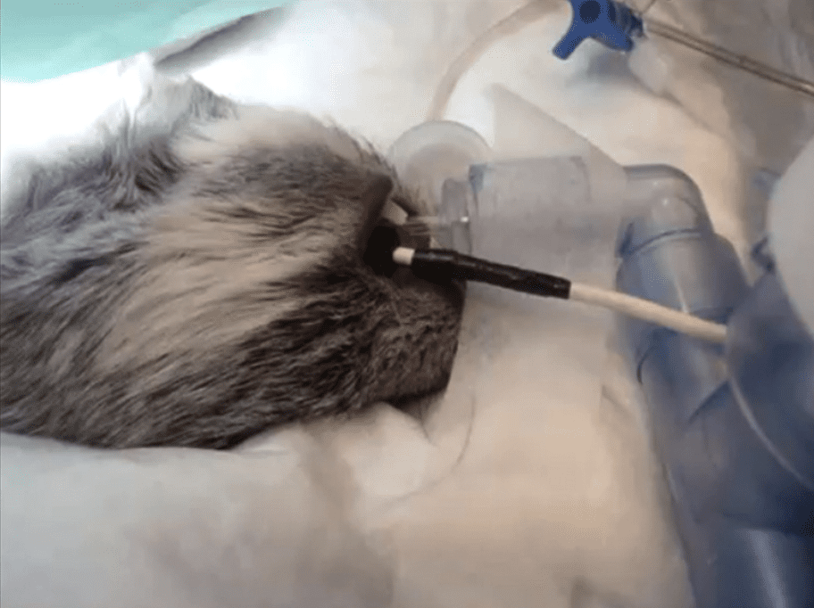
Bloat is the accumulation of gas in the stomach of degus. This accumulation can be related to poor nutrition, such as an excess of protein3 11. An unsuitable overall ration and slowed gut motility can contribute to gas accumulation. Water in fresh plants alone does not establish a mechanism for fatal protein-foam bloat in degus. Bloat can also occur due to slowed digestion, allowing the accumulation of bacteria and stomach fermentation. Dental malocclusions can also favor bloat when mouth breathing occurs2.
Degus show signs of abdominal pain by excessively “rubbing” their belly. They may eat less and become less active. It is crucial to ensure they eat and drink properly, and to obtain urgent veterinary assessment. A recovery diet may be prescribed after obstruction and other contraindications to oral feeding have been assessed.
Other Digestive Problems
Degus can develop other digestive issues, often related to poor nutrition, respiratory problems, or dental pathologies.
Cataract & Diabetes
It is often said that degus are “naturally diabetic,” but this is not entirely true12. While they are particularly sensitive to sugar, this is due to their very low insulin activity compared to other mammals13 and they tend to develop cataracts very rapidly, which may not necessarily be linked to diabetes. With controlled nutrition, it is possible to prevent or limit the progression of these conditions in degus.
Cataract
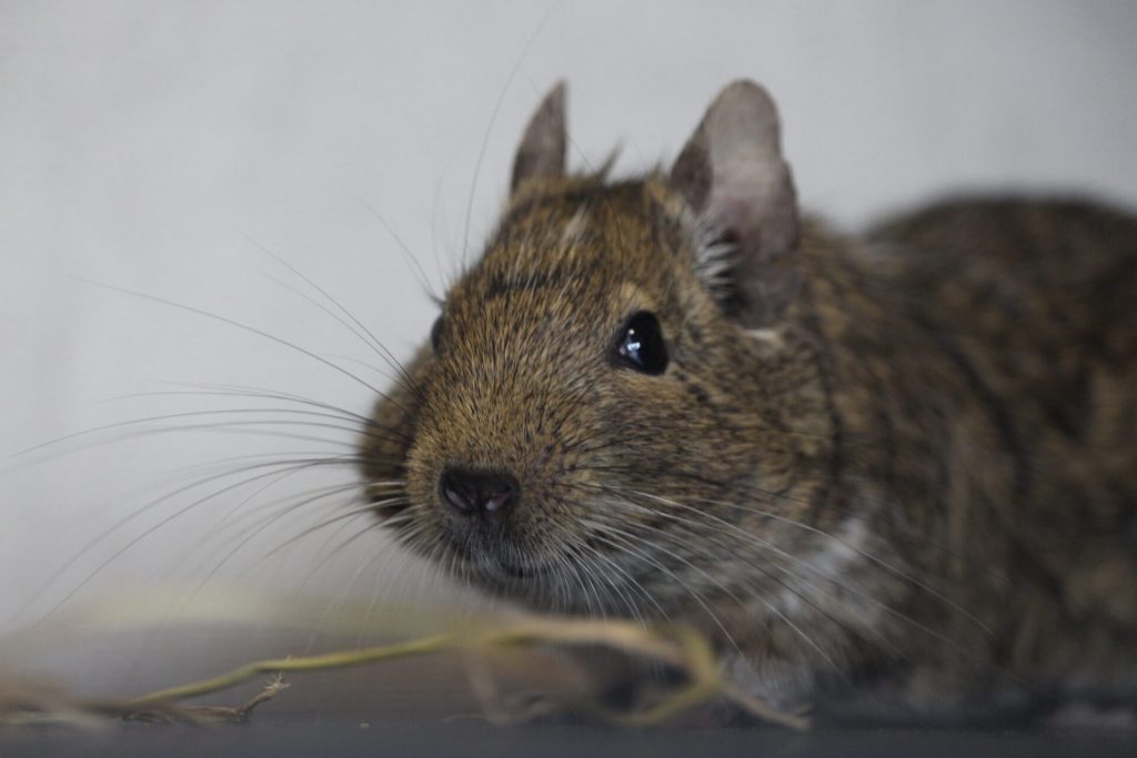
Cataract is a very common condition in degus14. It manifests as “whitening” of the eye, appearing as a spot that can eventually cover the entire eye. Degus may lose visual acuity and even go blind. Many captive degus can adapt to reduced vision, although the impact varies between individuals. Adapt the enclosure for safety and avoid frequent rearrangements.
Cataracts can be associated with diabetes, but they also occur in degus without confirmed diabetes. Their high sensitivity to sugar makes inappropriate nutrition a common cause of cataracts. Cataracts were recorded in approximately 13% of the animals in the retrospective clinical series2 especially in older degus who are more vulnerable to this condition.
Hyperinsulinemia
Degus can also develop hyperinsulinemia, which means having a high level of insulin (the hormone that regulates sugar) in the blood. To compensate for their low insulin activity, degus increase their insulin concentration and slow down its degradation when fed a high-carbohydrate diet. If degus are fed a diet high in carbohydrates, hyperinsulinemia can develop rapidly. Hyperinsulinemia often leads to the formation of cataracts and, if the pancreas is damaged, it can even result in diabetes. The symptoms of hyperinsulinemia are often not very visible but can lead to reduced activity, weight gain, and cataracts.
Hyperglycemia
Hyperglycemia can affect degus due to poor nutrition, especially if their diet consists of simple sugars. Hyperglycemia is generally episodic but can easily be confused with diabetes. If the pancreas is not damaged, it is often sufficient to change the degu’s diet to resolve hyperglycemia. However, if it persists, insulin therapy can be considered by a veterinarian.
The symptoms of hyperglycemia are similar to those of diabetes, including reduced activity, dizziness, difficulty moving, intense thirst, frequent urination, and the development of cataracts.
Diabetes
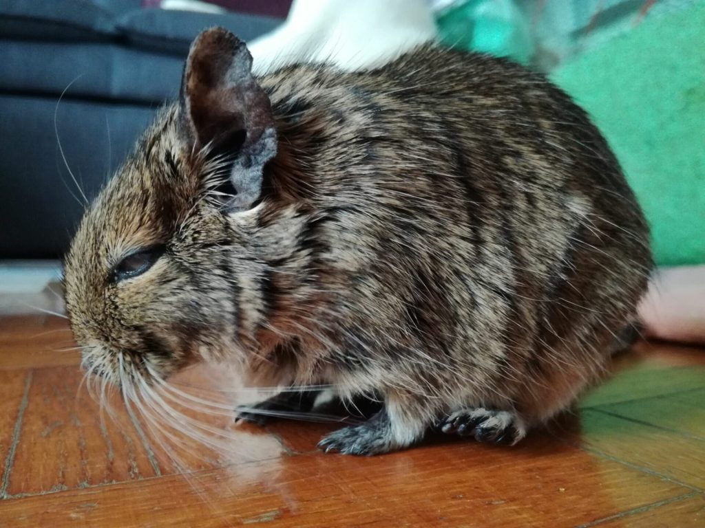
Diabetes mellitus is a condition that can be found in degus due to their lower insulin activity compared to other mammals15. If degus are fed an inappropriate diet, it can trigger diabetes. When degus have diabetes, it means they can no longer secrete insulin normally and cannot regulate blood sugar correctly. Consequently, blood sugar levels become abnormally high, leading to significant health problems, including cataracts, hepatic steatosis (excess fat in the liver), and hormone regulation disruptions.
Symptoms in degus are diverse and may include obesity, weight loss, anorexia, cataracts, dehydration, increased urination, excessive thirst, and gastrointestinal dysbiosis. Diagnosis requires appropriate testing and interpretation of persistent hyperglycaemia together with clinical findings; glycosuria is assessed in urine, not in a blood test, and hypercholesterolaemia is not a mandatory diagnostic criterion. Management includes dietary correction and, in selected cases, carefully monitored medication such as insulin prescribed by the veterinarian. However, it is important not to confuse hyperglycemia or hyperinsulinemia with diabetes, which was recorded in approximately 4% of the animals in that clinical series.
Skin Issues & Dermatological Problems
Skin problems are common in degus and can have various causes, such as excessive grooming, injuries, or parasites3.
Abscesses & Wounds
Skin problems, including abscesses and wounds, are common in degus2; injuries are often linked to fights between individuals, with injuries commonly occurring around the ears, head, and tail. It is essential to thoroughly disinfect the wound and promptly consult a veterinarian if it is substantial to prevent the development of abscesses. Antibiotic treatment may be considered, along with abscess cleaning and drainage if necessary.
Excessive Grooming
Excessive grooming is a common behavior in this species. It can be self-grooming or directed towards one or more companions. This behavior is typically associated with intense stress or pain. Identifying the underlying cause is crucial to stopping or reducing this behavior16. Common reasons include social isolation, a lack of opportunities for foraging/occupation, a dysfunctional social group, an inadequate cage, or dental issues.
In cases of excessive self-grooming, degus may focus particularly on their front paws. When this behavior is directed at a conspecific, areas with hair loss can be observed around the eyes, on the dorsal spine, flanks, and lower back.
Dermatophytosis / Ringworm
Mycological (fungal) conditions are relatively rare in degus. Additionally, it is important to note that the most common ringworm in degus (Trichophyton mentagrophytes) is not fluorescent under a Wood’s lamp and is potentially transmissible to humans. Typically observed symptoms include hair loss and thickening of the skin. The head is the most affected area, but degus can also be affected in other areas.
Dermatitis
Bacterial or ectoparasitic dermatitis is relatively rare in degus17. However, degus can become infected with certain mites, such as Demodex or Ornithonyssus bacoti, some of which may also bite people. The resulting lesions are varied and may include redness, itching, flaking, swelling, oozing, crusts/blisters, and skin thickening.
Pododermatitis
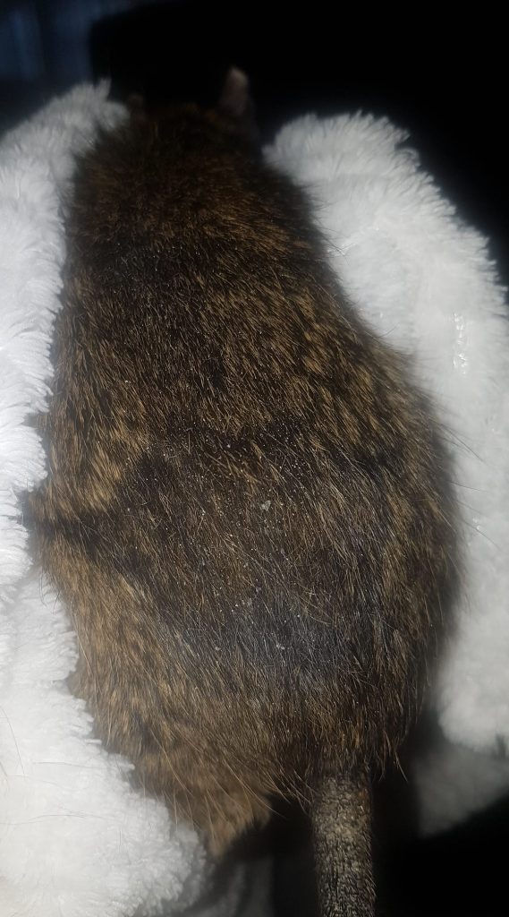
Pododermatitis is an infection resulting from repeated skin irritation on the paw pads. It can be associated with abscesses and inflammation, leading to significant lameness. Pododermatitis can be caused by hygiene issues as well as a lack of physical activity and obesity.
Parasites
Degus can be affected by various parasites, such as fleas, ticks, or mites, which cause skin bites or hair loss. It is important to treat both the degus and their environment to eliminate these parasites, which can persist in fabrics, bedding or hay.
Tail Loss/Scalping
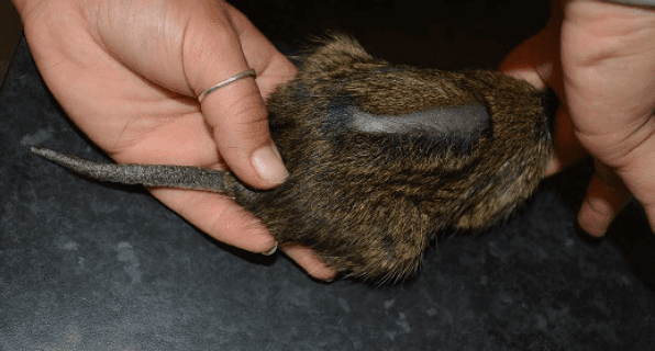
The tail skin can tear away after gripping, pulling, fighting or entrapment. This traumatic degloving is called tail slip or false caudal autotomy, and the tail does not regrow. Exposed vertebrae and damaged tissues require prompt veterinary care, including pain relief, wound management and surgery when indicated. Never grip or lift a degu by the tail.
Respiratory Diseases
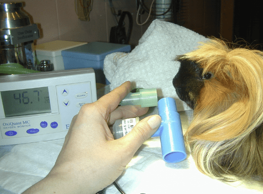
Respiratory diseases are observed in degus and are frequently associated with dental problems7 8. Infectious diseases remain rare, but rhinitis is common in cases of malocclusion. When a respiratory issue is observed in degus, it is important to diagnose it through cranial and thoracic imaging. The treatment then depends on the cause of the respiratory problem and the severity of the condition.
Reproductive Organ Pathologies
Some disorders can affect the reproductive organs of the degu, although these conditions are generally rare. The most common is penile prolapse in males.
Male
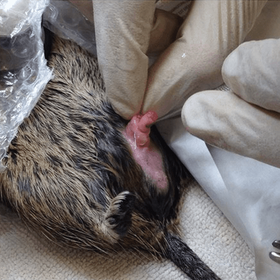
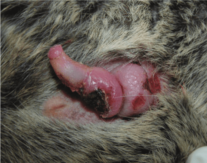
Penile prolapse and paraphimosis are conditions that prevent the retraction of the penis into its sheath and can occur in male degus. These problems are observed in case of injury or trauma, excessive mating, a constricting hair ring, or dirt accumulation around the penis. If the penis is not returned to its normal position, an ulcer can develop, as well as inflammation or even necrosis. The latter can lead to a blockage of the urethra and urinary tract disorders. Therefore, it is important to consult a veterinarian promptly if the organ does not return to its normal position. Amputation may be considered if the veterinarian is unable to retract the degu’s penis18.
Female
Female degus may experience some genital issues, such as dystocia (complications during childbirth), genital hematomas, pyometra (accumulation of pus in the uterus), or vaginal tumors1.
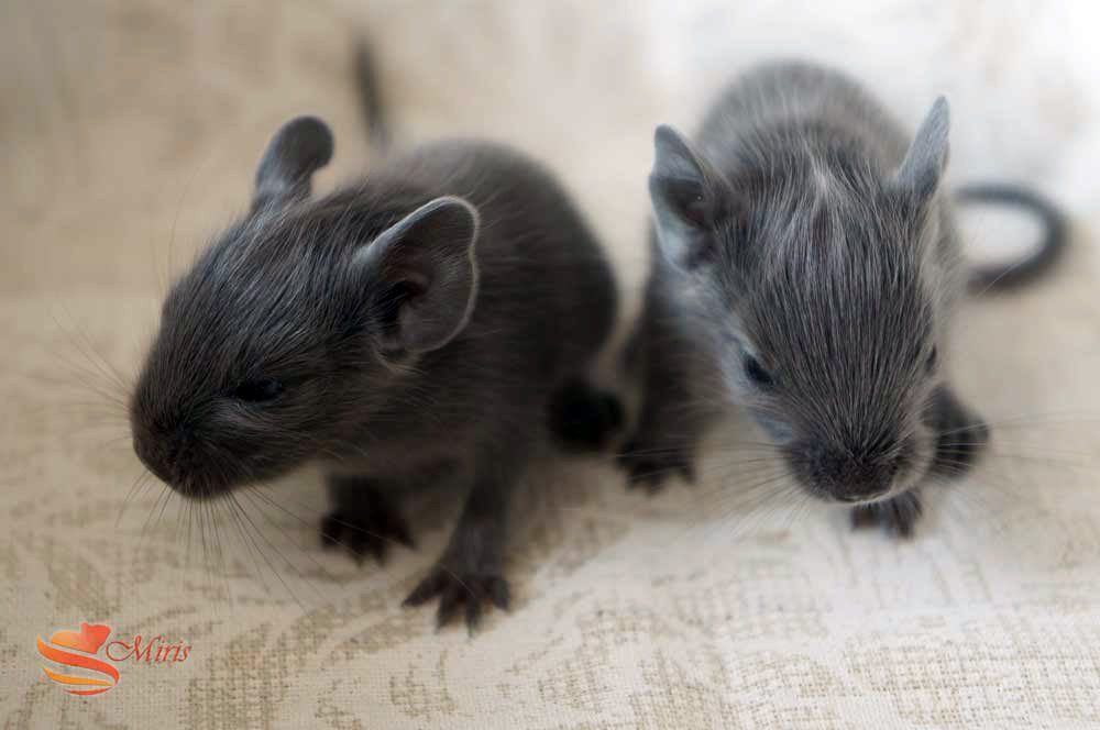
Difficult birth may involve oversized or malpositioned pups, obstruction or inadequate uterine contractions. Maternal health and body condition can also matter. Prolonged straining without progress, weakness, anorexia, rapid breathing, abdominal swelling or abnormal bleeding require urgent veterinary advice; do not wait for a fixed interval if the female is distressed.
Cancer and other tumours
Degus can develop benign and malignant tumours. Published case series document several types, but their frequency in the general pet population is uncertain. Elodontomas are proliferative dental lesions and must not automatically be labelled malignant cancers or described as inevitably becoming cancer.
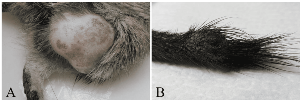 A: Fibrosarcoma, B: Chordoma
Several types of cancers are found in degus20 21 :
- Lung cancer: Octodon degus can develop a cancer similar to “smoker’s cancer,” such as bronchioloalveolar carcinoma, which was diagnosed in an adult male in a zoo.
- Kidney cancer: Renal and hepatic metastases have been found in the same individual; however, it appears that the behaviour of a tumour depends on its type and grade; some degu cancers metastasise.
- Lymphoma and other round-cell tumours: A few cases of cancerous masses related to lymphoma cancer (histiocytic lymphoma) have been found. In one degu, this condition caused death because one of the lymph nodes compressed the trachea.
- Liver cancer: Two degus developed liver cancer, specifically hepatocellular carcinoma. Unfortunately, it seems that this cancer metastasizes, as tumors affected the gallbladder, heart, and lungs. One of these degus also developed cholangiocarcinoma, a cancer of the bile duct cells.
- Proliferative dental lesions (elodontoma/odontoma): These are not automatically malignant cancers and may be associated with abnormal dental tissue growth10.
- Tail cancer: Tail cancer, or chordomas, is observed in degus. The tumor often appears as a mass on the tail and can be mistaken for an abscess.
Renal Disease
Octodon degus can be affected by several renal diseases, including nephrosis, pyelonephritis (infection), glomerulonephritis, renal polycystic disease, and renal neoplasms17. Symptoms vary widely and may include anorexia, weight loss, and a hunched posture.
Musculoskeletal Disorders
Degus may experience musculoskeletal disorders, with the most common ones being fractures, arthritis/spondylosis. Cases of bacterial arthritis, osteosarcoma (cancers), and fibrosarcoma (cancer) have also been observed.
Arthritis
Conditions like arthritis should be assessed promptly to manage pain and limit deterioration in mobility in degus22.
Octodon degus suffering from arthritis – Video on Rongeurs.net
Fracture
Fractures are often related to falls or mishandling and can be treated in octodons. Healing time depends on the fracture, age, stability and treatment. Degus have small, delicate bones, which can complicate surgical repair; ten days is not a universal healing time. Pain relief, appropriate stabilisation and follow-up are essential.
Alzheimer’s-like changes
Elderly octodons can develop amyloid deposits in blood vessels and brain tissues that are similar to those observed in humans with Alzheimer’s disease23. They can also, like humans, show cognitive deficits, anxiety, and similar circadian rhythms. These research findings vary between populations and do not establish that every elderly pet degu develops Alzheimer’s disease. New neurological signs require veterinary assessment for other treatable causes as well.
Heatstroke
Heatstroke can be very serious in degus, which do not tolerate heat well, unlike cooler temperatures. Heatstroke is an emergency and can progress rapidly. Move the animal out of the heat and contact a veterinarian immediately for guidance on safe cooling and transport; do not force oral fluids into a distressed animal.
Stroke
Older degus can suffer cerebrovascular events, leading to a loss of mobility or uncontrolled behaviors, sometimes resembling epileptic seizures. It is important to visit a veterinarian as quickly as possible in such cases.
Other
Degus can experience some other issues, such as ear infections or torticollis, which may be related to dental malocclusions. Finally, in the wild, degus can asymptomatically contract the protozoan parasite Trypanosoma cruzi, responsible for Chagas disease.
Internal parasites are another possible problem. Outdoor exposure and foraging history may guide veterinary investigation, but feeding fresh plants alone does not justify routine deworming. Treatment should be based on exposure, clinical findings and appropriate testing.
Epilepsy has also been observed in some individuals but remains a very rare condition.
Long-term health
Degus can live long, healthy lives with appropriate care. However, its diet and lifestyle play a significant role in preventing the most common health problems, such as dental and dermatological issues. Therefore, it is essential to provide an environment suitable for the species to maintain good health throughout its life. This includes providing appropriate nutrition, including fiber and calcium, a sufficiently spacious cage, a stable social group, and adequate physical and mental activities.
Sources
Visticot, M.-E. C. (2002). Un nouvel animal de compagnie : l’octodon, Octodon degus. Veterinary doctoral thesis, École nationale vétérinaire d’Alfort. https://theses.vet-alfort.fr/telecharger.php?id=340
Jekl, V., Hauptman, K., Knotek, Z. (2011). Diseases in pet degus: a retrospective study in 300 animals. J Small Anim Pract, 52(2), 107-112. https://doi.org/10.1111/j.1748-5827.2010.01028.x
Kubiak, M. (Ed.) (2021). Handbook of Exotic Pet Medicine. Wiley Blackwell. https://doi.org/10.1002/9781119389934
Jekl, V., et al. (2011). Impact of pelleted diets with different mineral compositions on the crown size of mandibular cheek teeth and mandibular relative density in degus (Octodon degus). Vet Rec, 168(24), 641. https://doi.org/10.1136/vr.d2012
Gumpenberger, M., et al. (2012). Impact of a high-phosphorus diet on the sonographic and CT appearance of kidneys in degus, and possible concurrence with dental problems. Vet Rec, 170(6), 153. https://doi.org/10.1136/vr.100257
Mans, C. & Jekl, V. (2016). Anatomy and Disorders of the Oral Cavity of Chinchillas and Degus. Vet Clin North Am Exot Anim Pract, 19(3), 843-869. https://doi.org/10.1016/j.cvex.2016.04.007
Ardiaca García, M., et al. (2021). Respiratory Diseases in Guinea Pigs, Chinchillas and Degus. Veterinary Clinics of North America: Exotic Animal Practice, 24(2), 419-457. https://doi.org/10.1016/j.cvex.2021.02.001
Suckow, M. A., Stevens, K. A. & Wilson, R. P. (Eds.) (2012). The Laboratory Rabbit, Guinea Pig, Hamster, and Other Rodents. Academic Press. https://doi.org/10.1016/C2009-0-30495-X
Crossley, D. A. (2001). Dental disease in chinchillas in the UK. Journal of Small Animal Practice, 42(1), 12-19. https://doi.org/10.1111/j.1748-5827.2001.tb01977.x
Jekl, V., et al. (2008). Elodontoma in a Degu (Octodon degus). Journal of Exotic Pet Medicine, 17(3), 216-220. https://doi.org/10.1053/j.jepm.2008.04.001
Quesenberry, K. E., et al. (Eds.) (2021). Ferrets, Rabbits, and Rodents: Clinical Medicine and Surgery (4th ed.). Elsevier/Saunders. https://evolve.elsevier.com/cs/product/9780323484350?role=faculty
Edwards, M. S. (2009). Nutrition and Behavior of Degus (Octodon degus). Veterinary Clinics of North America: Exotic Animal Practice, 12(2), 237-253. https://doi.org/10.1016/j.cvex.2009.01.003
Ardiles, A. O., et al. (2013). Octodon degus (Molina 1782): a model in comparative biology and biomedicine. Cold Spring Harb Protoc, 2013(4), 312-318. https://doi.org/10.1101/pdb.emo071357
Kiziltoprak, H., et al. (2019). Cataract in diabetes mellitus. World J Diabetes, 10(3), 140-153. https://doi.org/10.4239/wjd.v10.i3.140
Fox, J. G. & Murphy, J. C. (1979). Cytomegalic virus-associated insulitis in diabetic Octodon degus. Vet Pathol, 16(5), 625-628. https://doi.org/10.1177/030098587901600520
Zoopsy (n.d.). Dermatologie et troubles comportementaux : une démarche globale. Veterinary information sheet. https://www.zoopsy.com/fiches_conseils/comportement-animal40-dermatologie-et-troubles-comportementaux-une-demarche-globale-schwobthaler.pdf
Richardson, V. C. G. (2003). Diseases of Small Domestic Rodents. Blackwell Publishing. https://doi.org/10.1002/9780470690840
Calvo Carrasco, D. & Abou‐Zahr, T. (2015). Surgical narrowing of the preputial orifice for treatment of recurrent penile prolapse in a degu (
Octodon degus
). Veterinary Record Case Reports, 3(1), e 000223. https://doi.org/10.1136/vetreccr-2015-000223
Banks, R. E., et al. (2016). Exotic Small Mammal Care and Husbandry. https://doi.org/10.1002/9781119265405
Švara, T., et al. (2020). Spontaneous Tumors and Non-Neoplastic Proliferative Lesions in Pet Degus (Octodon degus). Vet Sci, 7(1), E 32. https://doi.org/10.3390/vetsci7010032
Murphy, J. C., et al. (1980). Spontaneous lesions in the degu. In R.J.Montali&G.Migaki(Eds.), The Comparative Pathology of Zoo Animals,437–444. Smithsonian Institution Press. https://www.researchgate.net/publication/284508331_Spontaneous_lesions_in_the_degu
Rannou, F., Sellam, J. & Berenbaum, F. (2010). Physiopathologie de l’arthrose : conceptions actuelles. La Presse Médicale, 39(11), 1159-1163. https://doi.org/10.1016/j.lpm.2010.09.001
Inestrosa, N. C., et al. (2005). Human-like rodent amyloid-beta-peptide determines Alzheimer pathology in aged wild-type Octodon degu. Neurobiol Aging, 26(7), 1023-1028. https://doi.org/10.1016/j.neurobiolaging.2004.09.016