
Some photographs show disease or injury. These images are placed inside labelled expandable sections, so the article can be read without opening them.
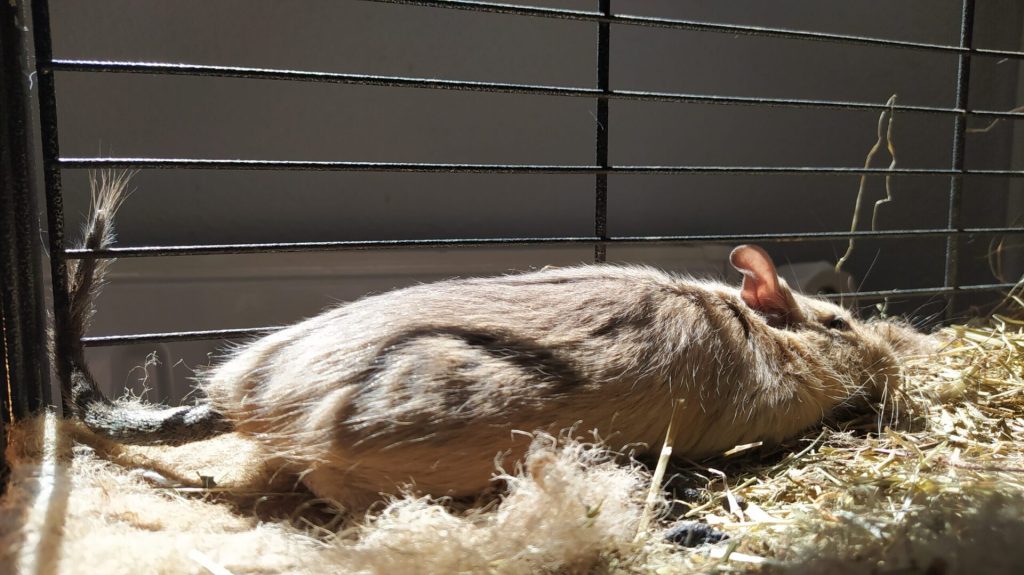
Degus can develop benign tumours, malignant tumours and non-neoplastic masses. Published information is much more limited than for common laboratory rats and mice, and differences between study populations prevent a simple comparison of overall cancer risk.1 Dental disease, diabetes, cataracts and skin disorders are also important clinical problems in this species. Dental swellings, including lesions called odontomas or elodontomas, must not automatically be described as cancer or as precursors that inevitably become malignant.2 An appropriate diet supports dental health, but cannot guarantee cancer prevention.
Diagnosis and treatment require a veterinarian familiar with degus. A lump’s appearance alone cannot reliably distinguish an abscess, dental lesion, benign tumour or cancer.

Tumours, cancer and other masses
A neoplasm is an abnormal growth of cells. Benign tumours do not invade and metastasise in the way malignant tumours do, but their size or location may still impair an organ and cause serious or even fatal complications. Malignant neoplasms are cancers; some form solid tumours, while others affect blood-forming or lymphoid tissues.
Non-neoplastic masses can resemble tumours but arise through other processes, including inflammation, tissue repair or other proliferative changes. Non-neoplastic therefore does not mean that no cells are multiplying; it describes the nature of the process.3
Cancer has a long evolutionary history. A remarkable example is an osteosarcoma diagnosed in the approximately 77-million-year-old dinosaur Centrosaurus apertus; this was not the oldest cancer ever identified.4 Veterinary oncology is well developed for dogs and cats,56 while degu-specific treatment evidence is limited. French rules governing anticancer medicines address safe handling and exposure; they should not be interpreted as a blanket ban on cancer treatment in rodents.7
How frequently are tumours reported?
The original article cites six tumour cases among 300 pet degus examined in one clinical series,2 proliferative lesions in 16 of 100 degus in another,8 and seven neoplasms among 189 animals in an older pathological report.9 These figures describe different selected populations and different outcomes. They cannot be averaged to produce a reliable prevalence for all degus, and do not support the earlier figure of 0.04%.
Age is relevant, and many reported patients were older animals.10 Nevertheless, a younger degu with a persistent lump should also be examined. Surgery is often considered when a localised lesion can be removed, but treatment decisions depend on its identity, location and the animal’s condition.
Possible contributing factors
The causes of most individual degu tumours are unknown. Factors discussed in wider cancer research include:
- Environment: exposure to pollutants and other potentially harmful agents, together with the broader effects of housing, stress and circadian disruption.
- Hormones: some mammary tumours in other mammals are hormonally influenced, but this does not establish the cause of a particular degu tumour.
- Genetics: laboratory-rat studies show that tumour patterns vary with strain and other factors. The contribution of inherited susceptibility in pet degus remains poorly characterised.11
- Diet: studies of diet and tumour metabolism in rats, including soy-protein experiments and measurements of hepatic gluconeogenesis, do not directly show that a particular degu food causes or prevents cancer.1213 Suitable fibre, energy and mineral balance remain important for general and dental health.
- Infection: a historical human estimate attributed about 16% of cancers to infections, including viruses, bacteria and parasites—not to viruses alone.14 Research has also explored mouse mammary tumour-like viruses in human breast cancer.15 Neither observation establishes an infectious cause of cancer in degus.
Signs that need investigation
Weigh your degus regularly and become familiar with their normal body condition. Possible signs include:
- Unexplained weight loss
- Reduced activity, interest or energy
- Loss of appetite or dehydration
- Dental abnormalities or swelling around the mouth16
- Skin changes or hair loss
- A lump on the tail, ears, head, abdomen, mouth or skin
- Breathing difficulties or nasal signs
- Lameness or abnormal limb swelling
- Seizures
These signs are not specific to cancer. Seek prompt veterinary assessment rather than assuming a diagnosis.
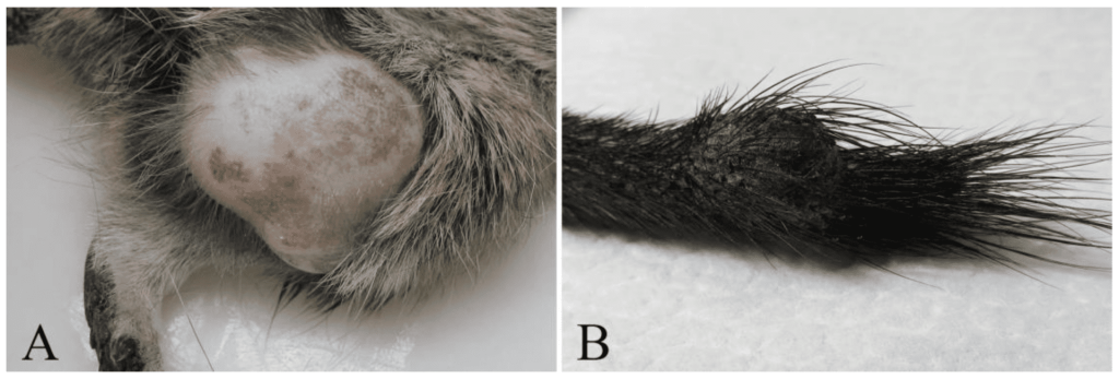
A: Fibrosarcoma. B: Chordoma.
Examination may be followed by sampling, cytology or biopsy, and imaging such as radiography or ultrasound. Histopathology is often needed to identify the lesion and assess its biological behaviour.
Treatment
Experimental chemotherapy has been studied in hamsters, rats and mice,1718 but these experiments are not validated treatment protocols for pet degus. The choice of treatment requires consideration of the diagnosis, likely benefit, administration, monitoring and quality of life. Where anticancer medicines are used, applicable safety requirements must also be followed.7
Surgical removal may be appropriate for a localised mass and sometimes requires removal of an affected digit or limb. Pain relief, supportive treatment and palliative care may also be important. It is too absolute to state that surgery is the only possible treatment in every case.
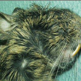
A mass associated with the incisor region. Its appearance alone cannot establish malignancy.
Examples of reported tumours
- Pulmonary carcinoma: bronchioloalveolar carcinoma has been reported in an adult male degu. This is a histological diagnosis, not proof of a smoking-related cause.
- Metastatic disease: renal and hepatic metastases have been described in a degu with pulmonary carcinoma. Secondary tumours in a kidney or liver should not be confused with a primary cancer of those organs.99
- Lymphoid and histiocytic tumours: older reports include lesions described using terms such as histiocytic lymphoma or reticulum-cell sarcoma. A cervical mass was reported to compress the trachea. Historical terminology may differ from modern classification.
- Hepatobiliary tumours: reports include hepatocellular carcinoma and cholangiocarcinoma, a malignancy of the bile-duct epithelium. Some cases involved additional organs, including the gallbladder, heart or lungs; the available reports do not justify a universal statement about the speed of spread.
- Oral and dental lesions: masses may be neoplastic or non-neoplastic. Elodontoma should not automatically be equated with an oral malignancy or assumed to undergo malignant transformation.16
- Tail chordoma: a firm tail mass can resemble an abscess externally and needs diagnostic assessment.19
An earlier literature review grouped reported tumours into eight epithelial, thirteen mesenchymal, four round-cell and two chordoma cases. These are counts from a collection of reports, not population prevalence.20 Other described lesions include splenic haemangioma, mesenteric lipoma, vaginal leiomyosarcoma, fibrosarcoma and renal transitional-cell carcinoma.
The limited literature shows that many tissues can be affected. A persistent mass needs individual investigation; the small and heterogeneous published series cannot establish a precise cancer rate for all captive degus.
Sources
Bomhard, E. (1992). Frequency of spontaneous tumors in Wistar rats in 30-months studies. Exp Toxicol Pathol, 44(7), 381-392. https://doi.org/10.1016/s0940-2993(11)80171-5
Jekl, V., Hauptman, K., Knotek, Z. (2011). Diseases in pet degus: a retrospective study in 300 animals. J Small Anim Pract, 52(2), 107-112. https://doi.org/10.1111/j.1748-5827.2010.01028.x
Shah, A., et al. (2020). Non-neoplastic Soft Tissue Tumors and Tumor-like Lesions. Semin Musculoskelet Radiol, 24(6), 645-666. https://doi.org/10.1055/s-0040-1713606
Ekhtiari, S., et al. (2020). First case of osteosarcoma in a dinosaur: a multimodal diagnosis. The Lancet Oncology, 21(8), 1021-1022. https://doi.org/10.1016/s1470-2045(20)30171-6
Vetopedia (n.d.). La chimiothérapie pour nos animaux de compagnie. https://www.vetopedia.fr/chimiotherapie-animaux-compagnie/
École nationale vétérinaire d’Alfort (n.d.). Cancérologie. Hôpital des animaux de compagnie. https://www.vet-alfort.fr/services-cliniques/hopital-des-animaux-de-compagnie/consultations-sur-rendez-vous/cancerologie-2
République française (2009). Arrêté du 18 juin 2009 relatif aux bonnes pratiques d’emploi des médicaments anticancéreux en médecine vétérinaire. Légifrance. https://www.legifrance.gouv.fr/loda/id/JORFTEXT000020764171/
Švara, T., et al. (2020). Spontaneous Tumors and Non-Neoplastic Proliferative Lesions in Pet Degus (Octodon degus). Vet Sci, 7(1), E32. https://doi.org/10.3390/vetsci7010032
Murphy, J. C., Crowell, T. P., Hewes, K. M., Fox, J. G., & Shalev, M. (1980). Spontaneous lesions in the degu. In The Comparative Pathology of Zoo Animals, pp. 437–444. Smithsonian Institution Press. https://www.researchgate.net/publication/284508331_Spontaneous_lesions_in_the_degu
Banks, R. E., et al. (2016). Exotic Small Mammal Care and Husbandry. https://doi.org/10.1002/9781119265405
Thompson, S. W., et al. (1961). Spontaneous tumors in the Sprague-Dawley rat. Journal of the National Cancer Institute, 27, 1037–1057. https://pubmed.ncbi.nlm.nih.gov/13920942/
Hakkak, R., & Korourian, S. (2020). Effects of soy protein diet on plasma isoflavones levels in obese DMBA-induced mammary tumor rat model. Cancer Research, 80(16 Supplement), abstract 4642. https://doi.org/10.1158/1538-7445.AM2020-4642
Liu, K. J., et al. (1990). Gluconeogenesis in the liver of tumor rats. J Surg Res, 49(2), 179-185. https://doi.org/10.1016/0022-4804(90)90259-5
Centre Léon Bérard (n.d.). Infections et cancer. Cancer Environnement. https://www.cancer-environnement.fr/fiches/expositions-environnementales/infections-et-cancer/
Lawson, J. S. & Glenn, W. K. (2019). Evidence for a causal role by mouse mammary tumour-like virus in human breast cancer. npj Breast Cancer, 5(1), 40. https://doi.org/10.1038/s41523-019-0136-4
Jekl, V., et al. (2008). Elodontoma in a Degu (Octodon degus). Journal of Exotic Pet Medicine, 17(3), 216-220. https://doi.org/10.1053/j.jepm.2008.04.001
Sugiura, K., Schmid, F. A., Brown, G. F., & González, R. (1971). Chemotherapy of hamster tumors. Cancer Chemotherapy Reports, Part 2, 2(1), 141–175. https://pubmed.ncbi.nlm.nih.gov/5565534/
Griswold, D. P., et al. (1974). Approaches to combination chemotherapy in rat, mouse, and hamster tumors. Cancer Chemotherapy Reports, Part 2, 4(1), 99–108. https://pubmed.ncbi.nlm.nih.gov/4524043/
Kondo, H., et al. (2018). CHORDOMA OF THE TAIL IN A DEGU (OCTODON DEGUS). Journal of Exotic Pet Medicine, 27(4), 1-4. https://doi.org/10.1053/j.jepm.2017.10.025
Sautier, L., et al. (2019). Coccygeal chordoma in a degu: case report and review of the literature. Journal of Veterinary Diagnostic Investigation, 31(1), 142-145. https://doi.org/10.1177/1040638718814584