
A vet experienced with degus must diagnose suspected diabetes and recommend treatment. Similar signs can have different causes, and self-diagnosis can delay appropriate care.
Degus are sometimes described as “naturally diabetic”. This is misleading: susceptibility to a disease does not mean that every animal has it. Degus can develop diabetes and cataracts, but these conditions must also be distinguished from hyperglycaemia and hyperinsulinaemia.
Blood glucose regulation and susceptibility
Insulin is a hormone involved in regulating the concentration of glucose in the blood. Caviomorph rodents, including degus, guinea pigs and chinchillas, have distinctive insulin sequences and biological properties. Experimental comparisons have reported lower insulin activity than in many other mammals, but this does not mean that healthy degus cannot regulate blood glucose.
Opazo and colleagues found fasting glucose and glucose-tolerance results within the expected mammalian range in the caviomorph rodents they examined, including degus. Compensatory mechanisms therefore need to be considered alongside insulin’s molecular properties. Higher hormone concentrations and changes in insulin handling have been discussed as possible adaptations; they should not automatically be labelled disease.
Degus nevertheless have a recognised susceptibility to disturbances of glucose regulation and associated cataracts. Pancreatic changes, including islet amyloidosis, have been described. A historical report also associated viral insulitis with diabetes, but this does not establish a viral cause for the usual pet case.
The role of diet
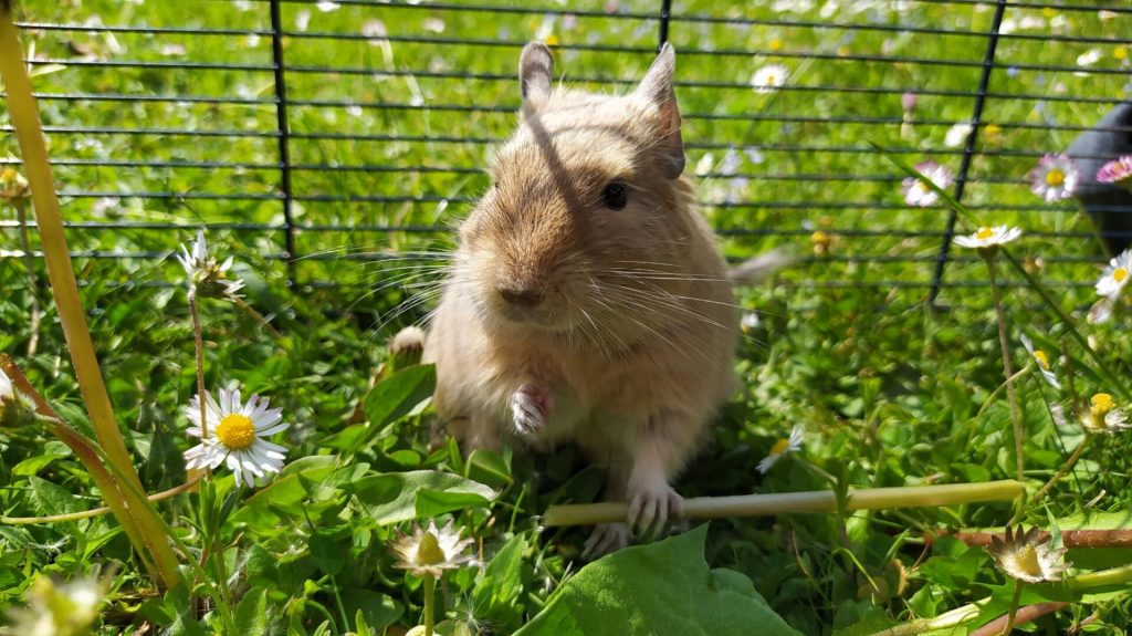
Diet is an important, modifiable part of care. Offer a fibre-rich diet, unrestricted suitable hay and a measured ration appropriate to the individual. Avoid sugary treats and fruit; do not treat “vegetables” as a single prohibited category, since suitable leafy vegetables can form part of an appropriate diet. Introduce changes carefully and assess the whole ration, including its starch content.
Age, body condition, inherited susceptibility and other illness also matter. Good feeding reduces avoidable risks but cannot guarantee that an animal will never develop diabetes or cataracts. See our feeding guide, foods and plants to avoid, and article on high-carbohydrate diets.
Conditions that should be distinguished
Cataracts
A cataract is an opacity of the lens inside the eye, rather than simply any white mark on its surface. It can reduce vision and may lead to blindness. Cataracts can occur with diabetes, but their presence alone does not diagnose diabetes: congenital, age-related and other causes must also be considered.
Cataracts were recorded in approximately 13.3% of the 300 patients in the cited veterinary series. This is a clinical sample, not an estimate of prevalence in all pet degus. Older animals may be affected, but cataracts are not restricted to old age.
Many visually impaired degus can adapt with suitable support. Keep essential resources accessible, prevent falls, maintain a familiar layout and monitor the animal’s ability to eat, drink and interact. It is too absolute to say that blindness never affects quality of life; assess the individual and seek veterinary advice.
Hyperinsulinaemia
Hyperinsulinaemia means an elevated blood insulin concentration. It is not interchangeable with diabetes or high blood glucose. In a species with unusual insulin properties, interpretation must take account of physiological compensation as well as possible insulin resistance and illness.
A high-carbohydrate diet can challenge glucose regulation, but a simple sequence in which excess insulin invariably causes cataracts and then pancreatic failure is not established by the cited evidence. Reduced activity, weight gain or a cataract cannot identify hyperinsulinaemia at home; a vet must interpret the relevant tests.
Hyperglycaemia
Hyperglycaemia means an elevated blood glucose concentration. Diet can contribute, but stress, illness and the conditions of sampling must also be considered. A transient reading is not automatically diabetes, and persistent hyperglycaemia must not be dismissed as a problem that only needs a diet change because the pancreas appears intact.
Possible warning signs include reduced activity, weakness or difficulty moving, marked thirst, increased urination and cataracts. These signs overlap with other conditions. Repeated testing and clinical assessment determine whether dietary management, treatment of an underlying problem or another intervention is required.
Diabetes mellitus
Diabetes mellitus involves persistently impaired glucose regulation associated with inadequate insulin secretion, action, or both. Glucose—not every form of carbohydrate—becomes abnormally elevated in the blood. Reported associated problems in degus include cataracts, fatty liver and broader metabolic or endocrine disturbances.
An affected animal may be overweight or may lose weight. Other possible signs include poor appetite, dehydration, increased urine production, excessive thirst, reduced activity and digestive disturbance. None is diagnostic on its own.
The vet assesses persistent hyperglycaemia alongside the history, examination and other tests. Glycosuria means glucose in the urine and is assessed through urinalysis; it is not a blood-test result. Blood lipid abnormalities may accompany metabolic disease, but hypercholesterolaemia is not a mandatory diagnostic criterion for diabetes. Approximately 4% of the animals in the cited clinical series were reported as diabetic; this must not be presented as the rate in the whole pet population.
Treatment and continuing care
Management is individual and requires veterinary supervision. Review the diet and body condition, investigate concurrent disease and monitor food and water intake, weight and clinical signs. Clean drinking water must remain available; see our water guide.
Insulin may be considered by the treating vet in selected cases. Administration and monitoring are demanding in such a small animal, and inappropriate insulin can cause dangerous hypoglycaemia. Do not give insulin or human diabetes medicines without a specific veterinary prescription and monitoring plan.
It is inaccurate to say that no continuing care is possible or that insulin is only relevant to an acute “crisis”. Equally, there is no universal treatment protocol that can be safely applied by owners. Cataracts require their own ophthalmic assessment and practical adaptations; changing the diet should not be promised to reverse an existing lens opacity.
Sources
Opazo, J. C., Soto-Gamboa, M., & Bozinovic, F. (2004). Blood glucose concentration in caviomorph rodents. Comparative Biochemistry and Physiology Part A: Molecular & Integrative Physiology, 137(1), 57–64. https://doi.org/10.1016/j.cbpb.2003.09.007
Opazo, J. C., Palma, R. E., Melo, F., & Lessa, E. P. (2005). Adaptive evolution of the insulin gene in caviomorph rodents. Molecular Biology and Evolution, 22(5), 1290–1298. https://doi.org/10.1093/molbev/msi117
Edwards, M. S. (2009). Nutrition and behavior of degus (Octodon degus). Veterinary Clinics of North America: Exotic Animal Practice, 12(2), 237–253. https://doi.org/10.1016/j.cvex.2009.01.003
Jekl, V., Hauptman, K., & Knotek, Z. (2011). Diseases in pet degus: a retrospective study in 300 animals. Journal of Small Animal Practice, 52(2), 107–112. https://doi.org/10.1111/j.1748-5827.2010.01028.x
Brown, C., & Donnelly, T. M. (2001). Cataracts and reduced fertility in degus (Octodon degus). Cataracts secondary to spontaneous diabetes mellitus. Lab Animal, 30(6), 25–26. https://pubmed.ncbi.nlm.nih.gov/11395944/
Nishi, M., & Steiner, D. F. (1990). Cloning of complementary DNAs encoding islet amyloid polypeptide, insulin, and glucagon precursors from a New World rodent, the degu, Octodon degus. Molecular Endocrinology, 4(8), 1192–1198. https://doi.org/10.1210/mend-4-8-1192
Fox, J. G., & Murphy, J. C. (1979). Cytomegalic virus-associated insulitis in diabetic Octodon degus. Veterinary Pathology, 16(5), 625–628. https://doi.org/10.1177/030098587901600520
Ardiles, A. O., et al. (2013). Octodon degus (Molina 1782): A model in comparative biology and biomedicine. Cold Spring Harbor Protocols, 2013(4), 312–318. https://doi.org/10.1101/pdb.emo071357