
Laboured or open-mouth breathing is an emergency. Contact a vet experienced with degus immediately, keep handling to a minimum and follow the clinic’s transport instructions. Do not force food or water into an animal struggling to breathe.
Degus can develop respiratory disease, including problems associated with dental disorders. They are related to guinea pigs and chinchillas within the broader rodent group, but belong to a different family, Octodontidae. Reports of comparatively fewer respiratory cases do not mean that degus are immune or that breathing changes can safely be ignored.

Possible causes
Dental disease and airway obstruction
In the clinical series cited by the original article, respiratory signs accounted for approximately 5.6% of 300 degu patients, while respiratory disease without another identified cause was reported at approximately 0.6%. These are observations from a veterinary patient group, not the proportion of all pet degus that will develop disease. Dental problems were an important association.
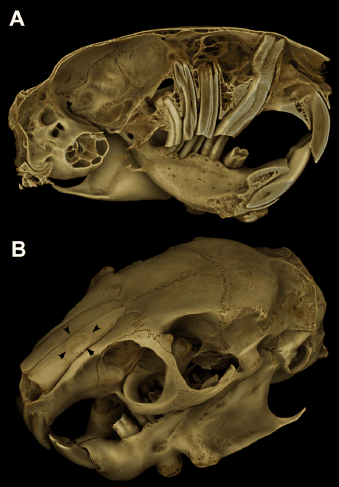
Elongated dental structures can encroach on the nasal cavity. An elodontoma is a proliferative dental lesion, not automatically a malignant cancer. A published degu case describes an elodontoma associated with severe dental disease and nasal obstruction. Other masses, foreign material or trauma after a fall or impact can also impair nasal airflow.
Other respiratory disorders
Rhinitis and bacterial infections are also considered. Organisms discussed in small-mammal respiratory medicine include Staphylococcus aureus, Streptococcus pneumoniae and Klebsiella pneumoniae. Their mention does not identify the pathogen in a particular degu, or establish that every species discussed in a review has the same susceptibility.
Environmental irritants or allergic disease may be considered after assessment. Remove obvious dust, smoke or fragrance exposures, but do not assume that respiratory distress is “just an allergy”. Cardiac disease and other causes may require different treatment.
Signs to recognise
Early changes can be subtle, and severe signs do not reveal the cause on their own. Watch for:
- Increased breathing effort: pronounced chest or abdominal movement, weakness or an unusual posture.
- Open-mouth breathing: a serious sign requiring immediate attention. Nasal obstruction is one possible cause, but not the only explanation for distress.
- Nasal discharge: clear or abnormal material around the nostrils, repeated nose wiping or reduced airflow. Trauma, dental disease and other disorders are possible; do not diagnose a simple “cold” or intestinal regurgitation from appearance alone.
- Unusual sounds: wheezing, whistling or crackling during breathing.
- Reduced activity or appetite: lethargy, refusal to move or eat, or a change in body temperature.
- Low blood oxygen: hypoxaemia is assessed clinically. The former “>92%” description was reversed and should not be used as a degu home-diagnostic threshold; measurement and interpretation depend on the patient and equipment.
- Eye or ear abnormalities: abnormal discharge or other concurrent signs can help the vet investigate the cause.
Veterinary diagnosis
Prompt assessment can limit suffering and identify a treatable underlying problem. Where dental disease is involved, both oral disease and the respiratory consequences need attention. Read the dental-health guide for the wider context.
The vet may use examination and auscultation, imaging, blood tests and appropriately selected microbiological or histopathological samples. Dental examination and endoscopy, radiographs or CT may be relevant where obstruction is suspected. An animal in respiratory distress may need stabilisation before stressful handling, anaesthesia or diagnostic procedures. These tests should be selected by the vet, rather than requested as a routine package for every breathing problem.
Treatment options
Treatment is chosen for the diagnosis, severity and individual risks—not simply a preference for a particular drug. The following describes options a clinician may consider, without providing a home-treatment protocol or doses. Management of the underlying cause remains essential.
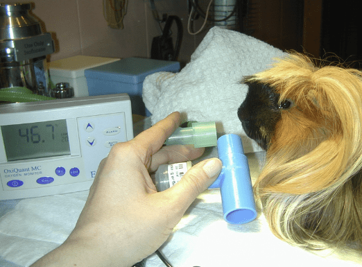
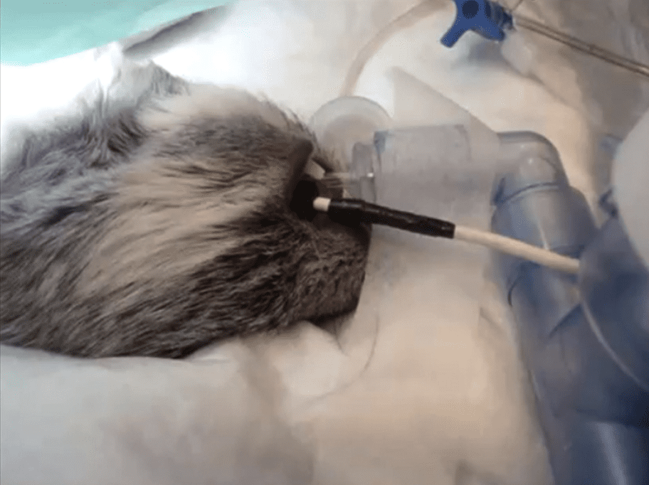
- Oxygen: a controlled delivery system, such as an oxygen enclosure or an appropriate mask, may support a hypoxaemic patient. The setup and response require monitoring.
- Stress reduction or carefully selected sedation: distress can increase oxygen demand, but sedative or anxiolytic drugs also carry risks in a compromised patient and require clinical supervision.
- Antibiotics: bacterial infection may require a species-appropriate antibiotic. Sampling, culture and susceptibility testing can guide the choice where feasible, without unnecessarily delaying urgent treatment.
- Corticosteroids: these are not a routine recommendation for degu pneumonia. Findings in human patients do not establish benefit in degus. The vet must weigh immunosuppression and metabolic effects, including hyperglycaemia, against a specific indication.
- Non-steroidal anti-inflammatory drugs: these may help pain or inflammation in selected patients, taking hydration, kidney function and other treatments into account.
- Bronchodilators: these act on airway smooth muscle and are useful only for particular indications. They do not resolve a nasal obstruction or every form of respiratory disease and can have adverse cardiovascular effects.
- Mucolytics: these alter mucus characteristics. Selection and administration require caution because evidence and tolerability vary; they should not be used indiscriminately.
- Antihistamines: a clinician may consider them for a justified allergic indication while also identifying and removing the trigger. Evidence in pet rodents is limited.
- Diuretics: these may be indicated for cardiogenic pulmonary oedema. Pleural effusion has several possible causes and may require drainage or other specific treatment; its presence does not automatically justify a diuretic. Dehydration and the underlying diagnosis matter.
- Nebulisation: a prescribed aerosol, sometimes involving sterile physiological saline, may support secretion management alongside other care. It is not the same as household steam or essential-oil inhalation.
- Nasal lavage: introducing fluid into the nasal passages is a clinical procedure with aspiration and handling risks. Do not attempt to flush a degu’s nose at home.
Seek veterinary advice at the first persistent breathing change, and urgent help for increased effort, open-mouth breathing, collapse or marked weakness. Supportive care cannot replace treatment of dental obstruction, infection, heart disease or another identified cause.
Sources
Ardiaca García, M., Montesinos Barceló, A., Bonvehí Nadeu, C., & Jekl, V. (2021). Respiratory Diseases in Guinea Pigs, Chinchillas and Degus. Veterinary Clinics of North America: Exotic Animal Practice, 24(2), 419–457. https://doi.org/10.1016/j.cvex.2021.02.001
Jekl, V., Hauptman, K., & Knotek, Z. (2011). Diseases in pet degus: a retrospective study in 300 animals. Journal of Small Animal Practice, 52(2), 107–112. https://doi.org/10.1111/j.1748-5827.2010.01028.x
Jekl, V., et al. (2008). Elodontoma in a Degu (Octodon degus). Journal of Exotic Pet Medicine, 17(3), 216–220. https://doi.org/10.1053/j.jepm.2008.04.001
Mans, C., & Jekl, V. (2016). Anatomy and Disorders of the Oral Cavity of Chinchillas and Degus. Veterinary Clinics of North America: Exotic Animal Practice, 19(3), 843–869. https://doi.org/10.1016/j.cvex.2016.04.007
Jekl, V., et al. (2011). Impact of pelleted diets with different mineral compositions on the crown size of mandibular cheek teeth and mandibular relative density in degus (Octodon degus). Veterinary Record, 168(24), 641. https://doi.org/10.1136/vr.d2012
Gumpenberger, M., et al. (2012). Impact of a high-phosphorus diet on the sonographic and CT appearance of kidneys in degus, and possible concurrence with dental problems. Veterinary Record, 170(6), 153. https://doi.org/10.1136/vr.100257
Edwards, M. S. (2009). Nutrition and behavior of degus (Octodon degus). Veterinary Clinics of North America: Exotic Animal Practice, 12(2), 237–253. https://doi.org/10.1016/j.cvex.2009.01.003
Colby, L. A., Rush, H. G., Mahoney, M. M., & Lee, T. M. (2012). Degu. In The Laboratory Rabbit, Guinea Pig, Hamster, and Other Rodents, pp. 1031–1053. Academic Press. https://www.sciencedirect.com/science/article/pii/B9780123809209000444